Lens (vertebrate anatomy)
The lens, or crystalline lens, is a transparent biconvex structure in most land vertebrate eyes. Along with the cornea, aqueous and vitreous humours it refracts light, focusing it onto the retina. This adjustment of the lens is known as accommodation (see also below). In many land animals the lens can accommodate by altering its shape to change the focal length of the eye allowing them to focus on objects at various distances. The accommodation allows a sharp real image of the object of interest to be formed on the retina. In many fully aquatic vertebrates such as fish other methods of accommodation are used such as changing the lens's position relative to the retina rather than changing lens shape. Accommodation is analogous to the focusing of a photographic camera via changing its lenses. In land vertebrates the lens is flatter on its anterior side than on its posterior side while fish the lens is often close to spherical.
| Lens | |
|---|---|
 Light from a single point of a distant object and light from a single point of a near object being brought to a focus by changing the curvature of the lens. | |
 Schematic diagram of the human eye. | |
| Details | |
| Part of | Eyeball |
| System | Visual system |
| Function | Refract light |
| Identifiers | |
| Latin | lens crystallin |
| MeSH | D007908 |
| TA98 | A15.2.05.001 |
| TA2 | 6798 |
| FMA | 58241 |
| Anatomical terminology | |
Accommodation in humans is well studied to allow artificial means of supplementing our focus such as glasses for correction of sight as we age. The refractive power of a younger human lens in its natural environment is approximately 18 dioptres, roughly one-third of the eye's total power of about 60 dioptres at infancy and 10 dioptres by the age of 25 years. Most of the reduction in our natural accommodation as we age is attributed to the aging of our lenses.
Structure
Position in the eye
The lens is located towards the front part of the vertebrate eye called the anterior segment which includes the cornea and iris positioned in front of the lens. The lens is held in place by the suspensory ligaments, attaching the lens at its equator to the rest of the eye[1][2] through the ciliary body. Behind the lens is the jelly like vitreous body which helps helps hold the lens in place. At the front of the lens is the liquid aqueous humor which bathes the lens with nutrients and other things. Land vertebrate lenses usually have an ellipsoid, biconvex shape. The front surface is less curved than the back. A human adult the lens is typically about 10mm in diameter and 4mm thick though changes shape with accommodation and size due to grow throughout a person's lifetime.[3]
Anatomy


The lens has three main parts: the lens capsule, the lens epithelium, and the lens fibers. The lens capsule is a relatively thick basement membrane forming the outermost layer of the lens. Inside the capsule much thinner lens fibers form the bulk of the lens. The cells of the lens epithelium form a thin layer between the lens capsule and the outermost layer of lens fibers at the front of the lens but not the back. The lens itself lacks nerves, blood vessels, or connective tissue.[4]
Lens capsule

The lens capsule is a smooth, transparent basement membrane that completely surrounds the lens. The capsule is elastic and its main structural component is collagen. It is synthesized by the lens epithelium and its main components in order of abundance are heparan sulfate proteoglycan (sulfated glycosaminoglycans (GAGs)), entactin, type IV collagen, laminin.[5] The capsule is very elastic and so allows the lens to assume a more spherical shape when not under the tension of the suspensory ligaments. The human capsule varies from 2 to 28 micrometres in thickness, being thickest near the equator (peri-equatorial region) and thinnest near the posterior pole.[3]
Lens epithelium
The lens epithelium is a single layer of cells at the front of the lens between the lens capsule and the lens fibers.[3] By providing the lens fibers with nutrients and removing waste the cells of the epithelium regulate maintain lens homeostasis.[6] As ions, nutrients, and liquid enter the lens from the aqueous humor, Na+/K+-ATPase pumps in the lens epithelial cells pump ions out of the lens to maintain appropriate lens osmotic concentration and volume, with equatorially positioned lens epithelium cells contributing most to this current. The activity of the Na+/K+-ATPases keeps water and current flowing through the lens from the poles and exiting through the equatorial regions.
The cells of the lens epithelium also divide into new lens fibers at the lens equator.[7] The lens lays down fibers from when it first forms in embryo until death.[8]
Lens fibers

The lens fibers form the bulk of the lens. They are long, thin, transparent cells, firmly packed, with diameters typically 4–7 micrometres and lengths of up to 12mm long in humans.[3] The lens fibers stretch lengthwise from the posterior to the anterior poles and, when cut horizontally, are arranged in concentric layers rather like the layers of an onion. If cut along the equator, it appears as a honeycomb. The middle of each fiber lies on the equator.[8] These tightly packed layers of lens fibers are referred to as laminae. The lens fiber cytoplasms are linked together via gap junctions, intercellular bridges and interdigitations of the cells that resemble "ball and socket" forms.
The lens is split into regions depending on the age of the lens fibers of a particular layer. Moving outwards from the central, oldest layer, the lens is split into an embryonic nucleus, the fetal nucleus, the adult nucleus, the inner and outer cortex. New lens fibers, generated from the lens epithelium, are added to the outer cortex. Mature lens fibers have no organelles or nuclei.
Development

Development of the vertebrate lens begins when the human embryo is about 4mm long. The accompanying picture shows the process in a more easily studied chicken embryo using plan english. Unlike the rest of the eye which is derived mostly from the inner embryo layers, the lens is derived from the skin around the embryo. The first stage of lens formation takes place when a sphere of cells formed by budding of the inner embryo layers comes close to the embyro's outer skin. The sphere of cells induces nearby outer skin to start changing into the lens placode. The lens placode is the first stage of transformation of a patch of skin into the lens. At this early stage, the lens placode is a single layer of cells.[9][10]
As development progresses, the lens placode begins to deepen and bow inwards. As the placode continues to deepen, the opening to the surface ectoderm constricts[11] and the lens cells bud off from the embryo's skin to form a sphere of cells known as the "lens vesicle". When the embryo is about 10mm long the lens vesicle has completely separated from the skin of the embryo.
The embryo then sends signals from the developing retina, inducing the cells closest to the posterior end of the lens vesicle to elongate toward the anterior end of the vesicle.[11] These signals also induce the synthesis of proteins called crystallins.[12] As the name suggests the crystallins can form a clear highly refractive jelly. These elongating cells eventually fill in the center of the vesicle with cells, that are long and thin like a strand of hair, called fibers. These primary fibers become the nucleus in the mature lens. The epithelial cells that don't form into fibers nearest the lens front give rise to the lens epithelium.
Additional fibers are derived from lens epithelial cells located at the lens equator. These cells lengthen towards the front and back wrapping around fibers already laid down. The new fibers need to be longer to cover earlier fibers but as the lens gets larger the ends of the newer fibers no longer reach as far towards the front and back of the lens. The lens fibers that do not reach the poles form tight, interdigitating seams with neighboring fibers. These seams being less crystalline then the bulk of the lens are more visible and are termed "sutures". The suture patterns become more complex as more layers of lens fibers are added to the outer portion of the lens.
The lens continues to grow after birth, with the new secondary fibers being added as outer layers. New lens fibers are generated from the equatorial cells of the lens epithelium, in a region referred to as the "germinative zone" and "bow region". The lens epithelial cells elongate, lose contact with the capsule and epithelium at the back and front of the lens, synthesize crystallin, and then finally lose their nuclei (enucleate) as they become mature lens fibers. In humans, as the lens grows by laying down more fibers through to early adulthood, the lens becomes more ellipsoid in shape. After about age 20 the lens grows rounder again and the iris is very important for this development.[3]
Several proteins control the embryonic development of the lens though PAX6 is considered the master regulator gene of this organ.[13] Other effectors of proper lens development include the Wnt signaling components BCL9 and Pygo2.[14]
Variations in lens structure
In many aquatic vertebrates, the lens is considerably thicker, almost spherical, to increase the refraction. This difference compensates for the smaller angle of refraction between the eye's cornea and the watery medium, as they have similar refractive indices.[15] Species that need to see well above and below water such as diving birds have the ability to change focus by 50 to 80 dioptres and have a somewhat altered lens and cornea structure and focus mechanisms to allow this.[16][17] Even among terrestrial animals the lens of primates such as humans is unusually flat going some way to explain why our vision is rather blurry under water.[18]
Function
Accommodation

The widely quoted Helmholtz mechanism of accommodation in the eye is often often referred to as a "model".[19] Direct experimental proof of any lens model is necessarily difficult as the vertebrate lens is transparent and only functions well in the living animals.
Helmholtz model
The lens is flexible and its curvature is thought to be controlled by ciliary muscles through the zonules. By changing the curvature of the lens, one can focus the eye on objects at different distances from it. This process is called accommodation, proposed Young in a lecture on 27th Nov 1800,[20] and refined by Helmholtz in 1909[21] is widely accepted.[22] At short focal distance the ciliary muscle contracts, zonule fibers loosen, and the lens thickens, resulting in a rounder shape and thus higher refractive power. Changing focus to an object at a greater distance requires the stretching of the lens by relaxing some of the sphincter like ciliary muscles while allowing the lens to be pulled thinner again so increasing the focal distance.

The Young model and intracapusular accommodation


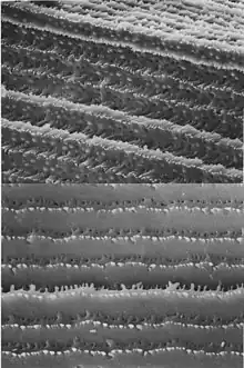
When Thomas Young proposed the changing of lens shape as the mechanism for focal accommodation in 1801 he thought the lens may be a muscle capable of contraction. This type of model is termed intracapsular accommodation. In a 1911 Nobel lecture Allvar Gullstrand spoke on "How I found the intracapsular mechanism of accommodation" and this aspect of lens focusing continues to be investigated.[23][24][25] Young spent time searching for the nerves that could stimulate the lens to contract without success. Since that time it has become clear the lens is not a simple muscle stimulated by a nerve so the 1909 Helmholtz model has taken a precedence. Neither Young, Helmholtz or Gullstrand had the benefit of many key twentieth century discoveries and techniques. Membrane proteins such as aquaporins which allow water to flow into and out of cells are the most abundant membrane protein in the lens.[26][27] Connexins which allow electrical coupling of cells are also prevalent. Electron microscopy and immunofluorescent microscopy show fiber cells to be highly variable in structure and composition.[28][29][30] Magnetic resonance imaging confirms a layering in the lens that may allow for different refractive plans within it.[31] The refractive index of human lens varies from approximately 1.406 in the central layers down to 1.386 in less dense layers of the lens.[32] This index gradient enhances the optical power of the lens. As more is learned about mammalian lens structure from in situ Scheimpflug photography, MRI[33][34] and physiological investigations it is becoming apparent the lens itself may not play only a passive role to the surrounding ciliary muscle but may be able to change its overall refractive index through mechanisms involving water dynamics in the lens still to be clarified.[35][36][37] The accompanying micrograph shows wrinkled fibers from a relaxed sheep lens indicating shortening of the lens fibers during near focus accommodation. The age related changes in the human lens may also be related to changes in the water dynamics in the lens.[38][39]
Lenses of birds, reptiles, amphibians and fish

In reptiles and birds, the ciliary body which supports the lens via suspensory ligaments also touches the lens with a number of pads on its inner surface. These pads compress and release the lens to modify its shape while focusing on objects at different distances; the suspensory ligaments usually perform this function in mammals. In fish and amphibians, the lens is fixed in shape, and focusing is instead achieved by moving the lens forwards or backwards within the eye.[18]
In cartilaginous fish, the suspensory ligaments are replaced by a membrane, including a small muscle at the underside of the lens. This muscle pulls the lens forward from its relaxed position when focusing on nearby objects. In teleosts, by contrast, a muscle projects from a vascular structure in the floor of the eye, called the falciform process, and serves to pull the lens backwards from the relaxed position to focus on distant objects. While amphibians move the lens forward, as do cartilaginous fish, the muscles involved are not similar in either type of animal. In frogs, there are two muscles, one above and one below the lens, while other amphibians have only the lower muscle.[18]
In the simplest vertebrates, the lampreys and hagfish, the lens is not attached to the outer surface of the eyeball at all. There is no aqueous humor in these fish, and the vitreous body simply presses the lens against the surface of the cornea. To focus its eyes, a lamprey flattens the cornea using muscles outside of the eye and pushes the lens backwards.[18]
Crystallins and transparency

Crystallins are water-soluble proteins that compose over 90% of the protein within the lens.[40] The three main crystallin types found in the human eye are α-, β-, and γ-crystallins. Crystallins tend to form soluble, high-molecular weight aggregates that pack tightly in lens fibers, thus increasing the index of refraction of the lens while maintaining its transparency. β and γ crystallins are found primarily in the lens, while subunits of α -crystallin have been isolated from other parts of the eye and the body. α-crystallin proteins belong to a larger superfamily of molecular chaperone proteins, and so it is believed that the crystallin proteins were evolutionarily recruited from chaperone proteins for optical purposes.[41] The chaperone functions of α-crystallin may also help maintain the lens proteins, which must last a human for their entire lifetime.[41]
Another important factor in maintaining the transparency of the lens is the absence of light-scattering organelles such as the nucleus, endoplasmic reticulum, and mitochondria within the mature lens fibers. Lens fibers also have a very extensive cytoskeleton that maintains the precise shape and packing of the lens fibers; disruptions/mutations in certain cytoskeletal elements can lead to the loss of transparency.[42]
The lens blocks most ultraviolet light in the wavelength range of 300–400 nm; shorter wavelengths are blocked by the cornea. The pigment responsible for blocking the light is 3-hydroxykynurenine glucoside, a product of tryptophan catabolism in the lens epithelium.[43] High intensity ultraviolet light can harm the retina, and artificial intraocular lenses are therefore manufactured to also block ultraviolet light.[44] People lacking a lens (a condition known as aphakia) perceive ultraviolet light as whitish blue or whitish-violet.[45][46]
Nourishment
The lens is metabolically active and requires nourishment in order to maintain its growth and transparency. Compared to other tissues in the eye, however, the lens has considerably lower energy demands.[47]
By nine weeks into human development, the lens is surrounded and nourished by a net of vessels, the tunica vasculosa lentis, which is derived from the hyaloid artery.[12] Beginning in the fourth month of development, the hyaloid artery and its related vasculature begin to atrophy and completely disappear by birth.[48] In the postnatal eye, Cloquet's canal marks the former location of the hyaloid artery.
After regression of the hyaloid artery, the lens receives all its nourishment from the aqueous humor. Nutrients diffuse in and waste diffuses out through a constant flow of fluid from the anterior/posterior poles of the lens and out of the equatorial regions, a dynamic that is maintained by the Na+/K+-ATPase pumps located in the equatorially positioned cells of the lens epithelium.[6]
Glucose is the primary energy source for the lens. As mature lens fibers do not have mitochondria, approximately 80% of the glucose is metabolized via anaerobic metabolism.[49] The remaining fraction of glucose is shunted primarily down the pentose phosphate pathway.[49] The lack of aerobic respiration means that the lens consumes very little oxygen as well.[49]
Clinical significance
- Cataracts are opacities of the lens. While some are small and do not require any treatment, others may be large enough to block light and obstruct vision. Cataracts usually develop as the aging lens becomes more and more opaque, but cataracts can also form congenitally or after injury to the lens. Nuclear sclerosis is a type of age-related cataract. Diabetes is another risk factor for cataract. Cataract surgery involves the removal of the lens and insertion of an artificial intraocular lens.
- Presbyopia is the age-related loss of accommodation, which is marked by the inability of the eye to focus on nearby objects. The exact mechanism is still unknown, but age-related changes in the hardness, shape, and size of the lens have all been linked to the condition.
- Ectopia lentis is the displacement of the lens from its normal position.
- Aphakia is the absence of the lens from the eye. Aphakia can be the result of surgery or injury, or it can be congenital.
Additional images
 MRI scan of human eye showing lens.
 Interior of anterior chamber of eye.
 The crystalline lens, hardened and divided.
 Section through the margin of the lens, showing the transition of the epithelium into the lens fibers.
 The structures of the eye labeled
 Another view of the eye and the structures of the eye labeled
 This svg file was configured so that the rays, diaphragm and crystalline lens are easily modified[50]
See also
- Accommodation reflex
- Crystallin
- Evolution of the eye, for how the lens evolved
- Intraocular lenses
- Iris
- Lens capsule
- Phacoemulsification
- Visual perception
- Zonules of Zinn
References
- "Equator of lens - definition from". Biology-Online.org. Archived from the original on 2012-03-22. Retrieved 2012-11-25.
- "equator of the crystalline lens - definition of equator of the crystalline lens in the Medical dictionary - by the Free Online Medical Dictionary, Thesaurus and Encyclopedia". Medical-dictionary.thefreedictionary.com. Retrieved 2012-11-25.
- John Forrester, Andrew Dick, Paul McMenamin, William Lee (1996). The Eye: Basic Sciences in Practice. London: W. B. Saunders Company Ltd. p. 28 ISBN 0-7020-1790-6
- Duker, Myron Yanoff, Jay S. (2008). Ophthalmology (3rd ed.). Edinburgh: Mosby. p. 382. ISBN 978-0323057516.
- Mohan, PS; Spiro, RG (25 March 1986). "Macromolecular organization of basement membranes. Characterization and comparison of glomerular basement membrane and lens capsule components by immunochemical and lectin affinity procedures". The Journal of Biological Chemistry. 261 (9): 4328–36. doi:10.1016/S0021-9258(17)35665-X. PMID 3512568.
- Candia, Oscar A. (2004). "Electrolyte and fluid transport across corneal, conjunctival and lens epithelia". Experimental Eye Research. 78 (3): 527–535. doi:10.1016/j.exer.2003.08.015. PMID 15106931.
- Shi, Yanrong; Barton, Kelly; De Maria, Alicia; Petrash, J. Mark; Shiels, Alan; Bassnett, Steven (15 May 2009). "The stratified syncytium of the vertebrate lens". Journal of Cell Science. 122 (10): 1607–1615. doi:10.1242/jcs.045203. PMC 2680101. PMID 19401333.
- "eye, human". Encyclopædia Britannica from Encyclopædia Britannica 2006 Ultimate Reference Suite DVD 2009
- Mitchell, PC (April 1891). "Double Chick Embryo". Journal of Anatomy and Physiology. 25 (Pt 3): 316–324.1. PMC 1328169. PMID 17231922.
- Chauhan, B; Plageman, T; Lou, M; Lang, R (2015). "Epithelial morphogenesis: the mouse eye as a model system". Current Topics in Developmental Biology. 111: 375–99. doi:10.1016/bs.ctdb.2014.11.011. PMC 6014593. PMID 25662266.
- Muccioli, Maria; Qaisi, Dalya; Herman, Ken; Plageman, Timothy F. (April 2016). "Lens placode planar cell polarity is dependent on Cdc42-mediated junctional contraction inhibition". Developmental Biology. 412 (1): 32–43. doi:10.1016/j.ydbio.2016.02.016. PMC 7370377. PMID 26902112.
- The Eye: Basic Sciences in Practice, p. 102, ISBN 0-7020-1790-6
- Cvekl, A.; Ashery-Padan, R. (2014). "The cellular and molecular mechanisms of vertebrate lens development". Development. 141 (23): 4432–4447. doi:10.1242/dev.107953. PMC 4302924. PMID 25406393.
- Cantù, Claudio; Zimmerli, Dario; Hausmann, George; Valenta, Tomas; Moor, Andreas; Aguet, Michel; Basler, Konrad (2014). "Pax6-dependent, but β-catenin-independent, function of Bcl9 proteins in mouse lens development". Genes & Development. 28 (17): 1879–1884. doi:10.1101/gad.246140.114. PMC 4197948. PMID 25184676.
- Kardong, K. (2008). Vertebrates: Comparative anatomy, function, evolution (5th ed.). (pp. 676–677). Boston: McGraw-Hill
- Katzir, Gadi; Howland, Howard C. (1 March 2003). "Corneal power and underwater accommodation in great cormorants( Phalacrocorax carbo sinensis )". Journal of Experimental Biology. 206 (5): 833–841. doi:10.1242/jeb.00142.
- Sivak, J.G.; Hildebrand, T.; Lebert, C. (January 1985). "Magnitude and rate of accommodation in diving and nondiving birds". Vision Research. 25 (7): 925–933. doi:10.1016/0042-6989(85)90203-2.
- Romer, Alfred Sherwood; Parsons, Thomas S. (1977). The Vertebrate Body. Philadelphia, PA: Holt-Saunders International. pp. 463–464. ISBN 978-0-03-910284-5.
- Land, Michael (19 April 2015). "Focusing by shape change in the lens of the eye: a commentary on Young (1801) 'On the mechanism of the eye'". Philosophical Transactions of the Royal Society B: Biological Sciences. 370 (1666): 20140308. doi:10.1098/rstb.2014.0308. PMC 4360117. PMID 25750232.
- Land, M (19 April 2015). "Focusing by shape change in the lens of the eye: a commentary on Young (1801) 'On the mechanism of the eye'". Philosophical Transactions of the Royal Society of London. Series B, Biological Sciences. 370 (1666). doi:10.1098/rstb.2014.0308. PMC 4360117. PMID 25750232.
- Helmholtz, H. von (1962). Treatise on physiological optics (English translation edited by JPC Southall. The Optical Society of America. From the third German Edition of Handbuch der Physiologischen Optik (1909), Leopold Voss, Leipzig. Dover reprint ed.). New York, NY: Dover Publications Inc.
- Fisher, R. F. (1 August 1977). "The force of contraction of the human ciliary muscle during accommodation". The Journal of Physiology. 270 (1): 51–74. doi:10.1113/jphysiol.1977.sp011938. PMC 1353417. PMID 915798.
- PAU, H (1952). "[Accommodative shift of the nucleus of the lens in intracapsular accommodation]". Klinische Monatsblatter fur Augenheilkunde und fur augenarztliche Fortbildung. 121 (2): 224–6. PMID 14955961.
- Huggert, Arne (27 May 2009). "The Intracapsular Mechanism of Accommodation". Acta Ophthalmologica. 42 (2): 389–397. doi:10.1111/j.1755-3768.1964.tb03627.x. PMID 14213923. S2CID 37325357.
- López-Gil, Norberto (3 March 2022). "Gullstrand Intracapsular Accommodation Mechanism Revised". Photonics. 9 (3): 152. Bibcode:2022Photo...9..152L. doi:10.3390/photonics9030152.
- Broekhuyse, R. M.; Kuhlmann, E. D.; Stols, A. L. (September 1976). "Lens membranes II. Isolation and characterization of the main intrinsic polypeptide (MIP) of bovine lens fiber membranes". Experimental Eye Research. 23 (3): 365–371. doi:10.1016/0014-4835(76)90135-4. PMID 976377.
- Mulders, SM; Preston, GM; Deen, PM; Guggino, WB; van Os, CH; Agre, P (14 April 1995). "Water channel properties of major intrinsic protein of lens". The Journal of Biological Chemistry. 270 (15): 9010–16. doi:10.1074/jbc.270.15.9010. PMID 7536742.
- Kuszak, J; Alcala, J; Maisel, H (December 1980). "The surface morphology of embryonic and adult chick lens-fiber cells". The American Journal of Anatomy. 159 (4): 395–410. doi:10.1002/aja.1001590406. PMID 7223675.
- Gruijters, WT; Kistler, J; Bullivant, S (October 1987). "Formation, distribution and dissociation of intercellular junctions in the lens". Journal of Cell Science. 88 ( Pt 3) (3): 351–9. doi:10.1242/jcs.88.3.351. PMID 3448099.
- Gruijters, WT (July 1989). "A non-connexon protein (MIP) is involved in eye lens gap-junction formation". Journal of Cell Science. 93 ( Pt 3) (3): 509–13. doi:10.1242/jcs.93.3.509. PMID 2691517.
- Hermans, EA; Dubbelman, M; Van der Heijde, R; Heethaar, RM (December 2008). "Equivalent refractive index of the human lens upon accommodative response". Optometry and Vision Science. 85 (12): 1179–84. doi:10.1097/OPX.0b013e31818e8d57. PMID 19050472. S2CID 205907383.
- Hecht, Eugene. Optics, 2nd ed. (1987), Addison Wesley, ISBN 0-201-11609-X. p. 178.
- Hermans, Erik A.; Pouwels, Petra J. W.; Dubbelman, Michiel; Kuijer, Joost P. A.; van der Heijde, Rob G. L.; Heethaar, Rob M. (1 January 2009). "Constant Volume of the Human Lens and Decrease in Surface Area of the Capsular Bag during Accommodation: An MRI and Scheimpflug Study". Investigative Ophthalmology & Visual Science. 50 (1): 281–289. doi:10.1167/iovs.08-2124. PMID 18676625.
- Stahnke, T.; Hadlich, S.; Wree, A.; Guthoff, R.; Stachs, O.; Langner, S. (16 December 2016). "Magnetresonanzmikroskopie des Akkommodationsapparats". Klinische Monatsblätter für Augenheilkunde. 233 (12): 1320–1323. doi:10.1055/s-0042-118599. PMID 27984837.
- Vaghefi, E; Pontre, BP; Jacobs, MD; Donaldson, PJ (August 2011). "Visualizing ocular lens fluid dynamics using MRI: manipulation of steady state water content and water fluxes". American Journal of Physiology. Regulatory, Integrative and Comparative Physiology. 301 (2): R335-42. doi:10.1152/ajpregu.00173.2011. PMID 21593426. S2CID 9525037.
- Donaldson, Paul J.; Chen, Yadi; Petrova, Rosica S.; Grey, Angus C.; Lim, Julie C. (December 2022). "Regulation of lens water content: Effects on the physiological optics of the lens". Progress in Retinal and Eye Research: 101152. doi:10.1016/j.preteyeres.2022.101152. PMID 36470825. S2CID 254243790.
- Suzuki-Kerr, H; Walker, KL; Han, MH; Lim, JC; Donaldson, PJ (2022). "Hyposmotic stress causes ATP release in a discrete zone within the outer cortex of rat lens". Molecular Vision. 28: 245–256. PMC 9514545. PMID 36284672.
- Moffat, BA; Landman, KA; Truscott, RJ; Sweeney, MH; Pope, JM (December 1999). "Age-related changes in the kinetics of water transport in normal human lenses". Experimental Eye Research. 69 (6): 663–9. doi:10.1006/exer.1999.0747. PMID 10620395.
- Jones, C.E.; Atchison, D.A.; Meder, R.; Pope, J.M. (August 2005). "Refractive index distribution and optical properties of the isolated human lens measured using magnetic resonance imaging (MRI)". Vision Research. 45 (18): 2352–2366. doi:10.1016/j.visres.2005.03.008. PMID 15979462. S2CID 8894700.
- Hoehenwarter, W.; Klose, J.; Jungblut, P. R. (2006). "Eye lens proteomics". Amino Acids. 30 (4): 369–389. doi:10.1007/s00726-005-0283-9. PMID 16583312. S2CID 19978371.
- Andley, Usha P. (2007). "Crystallins in the eye: Function and pathology". Progress in Retinal and Eye Research. 26 (1): 78–98. doi:10.1016/j.preteyeres.2006.10.003. PMID 17166758. S2CID 29317220.
- Bloemendal, Hans; De Jong, Wilfried; Jaenicke, Rainer; Lubsen, Nicolette H.; Slingsby, Christine; Tardieu, Annette (2004). "Ageing and vision: Structure, stability and function of lens crystallins". Progress in Biophysics and Molecular Biology. 86 (3): 407–485. doi:10.1016/j.pbiomolbio.2003.11.012. PMID 15302206.
- Andrew M.Wood and Roger J.W.Truscott (March 1993). "UV Filters in Human Lenses: Tryptophan Catabolism". Experimental Eye Research. 56 (3): 317–325. doi:10.1006/exer.1993.1041. PMID 8472787.
- Mainster, M. A. (2006). "Violet and blue light blocking intraocular lenses: Photoprotection versus photoreception". British Journal of Ophthalmology. 90 (6): 784–792. doi:10.1136/bjo.2005.086553. PMC 1860240. PMID 16714268.
- Anderson, Robert M. (1983). "Visual Perceptions and Observations of an Aphakic Surgeon". Perceptual and Motor Skills. 57 (3_suppl): 1211–1218. doi:10.2466/pms.1983.57.3f.1211. PMID 6664798. S2CID 20005737.
- Hambling, David (29 May 2002). "Let the light shine in". The Guardian.
- Whikehart, David R. (2003). Biochemistry of the Eye, 2nd ed. 2003. Philadelphia: Butterworth Heinemann, p. 107–8 ISBN 0-7506-7152-1
- The Eye: Basic Sciences in Practice, p. 104, ISBN 0-7020-1790-6
- Biochemistry of the Eye, 2nd ed, p. 107–8, ISBN 0-7506-7152-1
- Download and open with Inkscape 9.1. The separate components reside on different "layers" to facilitated editing.
External links
- Histology image: 08001loa – Histology Learning System at Boston University
Anatomy of the globe of the human eye | |||||||
|---|---|---|---|---|---|---|---|
| Fibrous tunic (outer) |
|  1:posterior segment 2:ora serrata 3:ciliary muscle 4:ciliary zonules 5:Schlemm's canal 6:pupil 7:anterior chamber 8:cornea 9:iris 10:lens cortex 11:lens nucleus 12:ciliary process 13:conjunctiva 14:inferior oblique muscule 15:inferior rectus muscule 16:medial rectus muscle 17:retinal arteries and veins 18:optic disc 19:dura mater 20:central retinal artery 21:central retinal vein 22:optic nerve 23:vorticose vein 24:bulbar sheath 25:macula 26:fovea 27:sclera 28:choroid 29:superior rectus muscle 30:retina | |||||
| Uvea / vascular tunic (middle) |
| ||||||
| Retina (inner) |
| ||||||
| Anatomical regions of the eye |
| ||||||
| Other |
| ||||||