Lacrimal nerve
The lacrimal nerve is the smallest branch of the ophthalmic nerve (CN V1), itself a branch of the trigeminal nerve (CN V). The other branches of the ophthalmic nerve are the frontal nerve and nasociliary nerve.[1]
| Lacrimal nerve | |
|---|---|
 Nerves of the orbit, the lacrimal nerve is visible, labelled over the eye. | |
| Details | |
| From | ophthalmic nerve |
| Innervates | lacrimal gland, conjunctiva, skin of lateral forehead, scalp and upper eyelid |
| Identifiers | |
| Latin | nervus lacrimalis |
| TA98 | A14.2.01.018 |
| TA2 | 6198 |
| FMA | 52628 |
| Anatomical terms of neuroanatomy | |
Structure
Origin
The lacrimal nerve branches from the ophthalmic nerve immediately before traveling through the superior orbital fissure to enter the orbit.
The lacrimal nerve receives a communicating branch from the zygomatic nerve which may carry some post-ganglionic parasympathetic nerve fibres from the pterygopalatine ganglion.[2]
Course
It enters the orbit through the superior orbital fissure outside the common tendinous ring, and lateral to the frontal nerve, and trochlear nerve (CN IV). Once inside the orbit, it travels anterior-ward along the lateral wall of the orbit upon the upper margin of the lateral rectus muscle.[2] It is accompanied by the lacrimal artery along its course through the orbit. It travels through the lacrimal gland,[2] supplying the gland with sensory and parasympathetic branches, then continuing anteriorly as a few small sensory branches.
Function
Sensory
The lacrimal nerve provides sensory innervation to the lacrimal gland, the skin and conjunctiva of the lateral portion of the upper eyelid[2] and superior fornix, and the skin of the lateral forehead and scalp.
Parasympathetic
The lacrimal nerve also provides parasympathetic innervation to the lacrimal gland from the communicating branch it receives from the zygomatic nerve.
Additional images
 Superior view of the nerves of the orbit. The lacrimal nerve is seen branching from the ophthalmic nerve.
 Sensory innervation to the skin of the head and neck. The cutaneous distribution of the lacrimal nerve can be seen above the eye in the green area.
 Anterior view of the orbit and tarsal plates. The lacrimal nerve can be seen exiting the orbit superolaterally after it supplies the lacrimal gland.
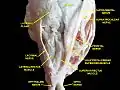 Superior view of a dissection of the left orbit. The lacrimal nerve is visible innervating the lacrimal gland.
References
- Gray's anatomy : the anatomical basis of clinical practice. Standring, Susan (41 ed.). [Philadelphia]. 2016. ISBN 978-0-7020-5230-9. OCLC 920806541.
{{cite book}}: CS1 maint: others (link) - Standring, Susan (2020). Gray's Anatomy: The Anatomical Basis of Clinical Practice (42nd ed.). New York. p. 782. ISBN 978-0-7020-7707-4. OCLC 1201341621.