Middle pharyngeal constrictor muscle
The middle pharyngeal constrictor is a fan-shaped muscle located in the neck. It is one of three pharyngeal constrictor muscles. It is smaller than the inferior pharyngeal constrictor muscle.
| Middle pharyngeal constrictor muscle | |
|---|---|
 | |
 Muscles of the pharynx, viewed from behind, together with the associated vessels and nerves (middle pharyngeal constrictor muscle labeled as Mid. constr. at center) | |
| Details | |
| Origin | Hyoid bone |
| Insertion | Pharyngeal raphe |
| Artery | Ascending pharyngeal artery |
| Nerve | Pharyngeal plexus of vagus nerve |
| Actions | Swallowing |
| Identifiers | |
| Latin | Musculus constrictor pharyngis medius |
| TA98 | A05.3.01.108 |
| TA2 | 2184 |
| FMA | 46622 |
| Anatomical terms of muscle | |
The middle pharyngeal constrictor originates from the greater cornu and lesser cornu of the hyoid bone, and the stylohyoid ligament. It inserts onto the pharyngeal raphe. It is innervated by a branch of the vagus nerve through the pharyngeal plexus. It acts to propel a bolus downwards along the pharynx towards the esophagus, facilitating swallowing.
Structure
The fibers diverge from their origin: the lower ones descend beneath the constrictor inferior, the middle fibers pass transversely, and the upper fibers ascend and overlap the constrictor superior.
Origin
The middle pharyngeal constrictor arises from the whole length of the upper border of the greater cornu of the hyoid bone, the lesser cornu of the hyoid bone, and from the stylohyoid ligament.
Insertion
The middle pharyngeal constrictor inserts posteriorly into the pharyngeal raphe, blending with its contralateral partner at the midline.
Innervation
Similarly to the superior and inferior pharyngeal constrictor muscles, it is innervated by a branch of the vagus nerve through the pharyngeal plexus.
Function
As soon as the bolus of food is received in the pharynx, the elevator muscles relax, the pharynx descends, and the constrictors contract upon the bolus, and convey it downward into the esophagus.[1][2] They also have respiratory mechanical effects.[3]
Additional images
 Hyoid bone. Anterior surface. Enlarged.
 Muscles of the neck. Lateral view.
 Middle pharyngeal constrictor muscle
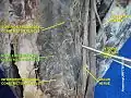 Middle pharyngeal constrictor muscle
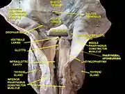 Deep dissection of larynx, pharynx and tongue seen from behind
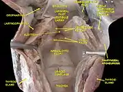 Deep dissection of larynx, pharynx and tongue seen from behind
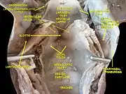 Deep dissection of larynx, pharynx and tongue seen from behind
References
![]() This article incorporates text in the public domain from page 1143 of the 20th edition of Gray's Anatomy (1918)
This article incorporates text in the public domain from page 1143 of the 20th edition of Gray's Anatomy (1918)
- Rowe LD, Miller AJ, Chierici G, Clendenning D (August 1984). "Adaptation in the function of pharyngeal constrictor muscles". Otolaryngology–Head and Neck Surgery. 92 (4): 392–401. doi:10.1177/019459988409200404. PMID 6435057. S2CID 32361287.
- Donner, Martin W.; Bosnia, James F.; Robertson, Diane L. (1985). "Anatomy and physiology of the pharynx". Gastrointestinal Radiology. 10 (1): 197–212. doi:10.1007/BF01893103. ISSN 0364-2356. PMID 4029536. S2CID 37515662.
- Kuna, Samuel T (2000). "Respiratory-related activation and mechanical effects of the pharyngeal constrictor muscles". Respiration Physiology. 119 (2–3): 155–161. doi:10.1016/S0034-5687(99)00110-3. ISSN 0034-5687. PMID 10722858.
Further reading
- Its role in speech: Hamaker, Ronald C.; Blom, Eric D. (2003). "Botulinum Neurotoxin for Pharyngeal Constrictor Muscle Spasm in Tracheoesophageal Voice Restoration". The Laryngoscope. 113 (9): 1479–1482. doi:10.1097/00005537-200309000-00010. ISSN 0023-852X. PMID 12972919. S2CID 12251825.
- Its role in Hyoid bone syndrome: Ernest, Edwin A.; Salter, E. George (1991). "Hyoid bone syndrome: A degenerative injury of the middle pharyngeal constrictor muscle with photomicroscopic evidence of insertion tendinosis". The Journal of Prosthetic Dentistry. 66 (1): 78–83. doi:10.1016/0022-3913(91)90357-3. ISSN 0022-3913. PMID 1941681.
External links
- lesson8 at The Anatomy Lesson by Wesley Norman (Georgetown University) (latpharyngealitems3)